Introduction
Every time you open your eyes, an extraordinary biological process begins. Understanding how your eyes turn light into vision starts with following the journey of light as it enters the eye, reaches the retina, and is converted into signals that the brain can interpret.
This process allows you to recognize faces, read words, appreciate colors, judge distances, and navigate your surroundings. Yet your eyes do not actually create the images you experience. They capture light and convert it into information that your brain must interpret.
Understanding how your eyes turn light into vision reveals a remarkable connection between optics, cellular biology, and the human nervous system.
How Does Light Enter the Eye?
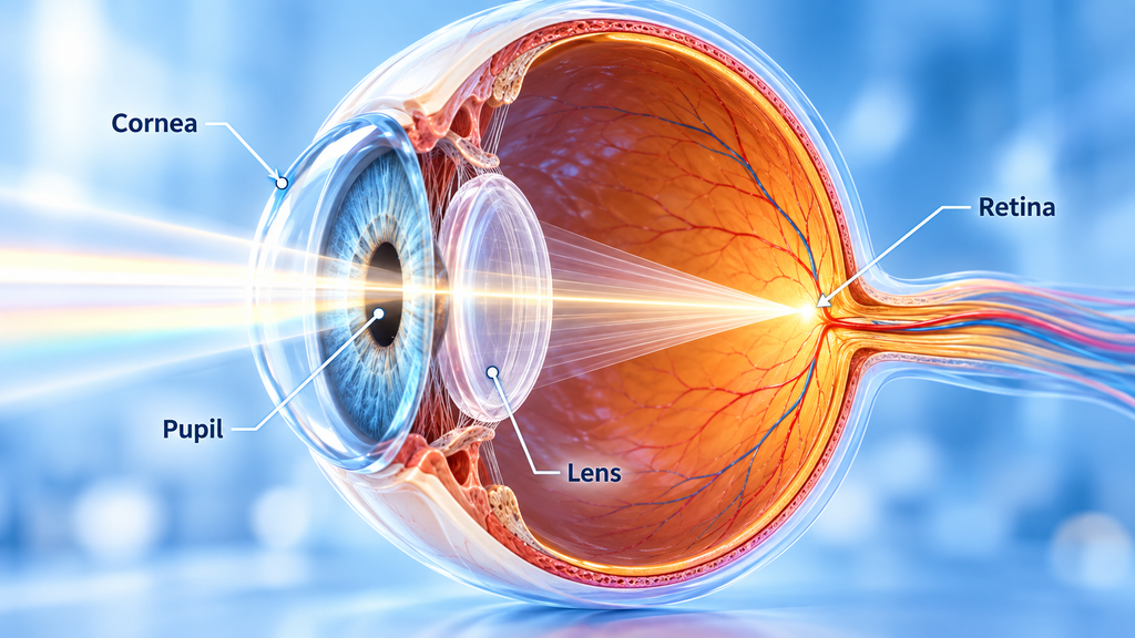
The cornea: The eye’s first focusing surface
Light first passes through the cornea, the transparent, dome-shaped structure covering the front of the eye.
The cornea bends incoming light through a process called refraction. It provides much of the eye’s focusing power, helping direct light toward the retina.
For clear vision, the cornea must maintain its transparency and proper curvature.
The iris and pupil: Controlling incoming light
After passing through the cornea, light travels through the pupil, the opening in the center of the iris.
The iris is the colored part of your eye. Its muscles adjust pupil size according to lighting conditions.
In bright environments, the pupil becomes smaller to limit incoming light. In darkness, it widens to allow more light to enter.
The lens: Fine-tuning the focus
Behind the iris is the crystalline lens, a transparent and flexible structure that adjusts the eye’s focus.
When you look at a nearby object, the ciliary muscles contract, allowing the lens to become rounder. For distant objects, the lens becomes flatter.
This adjustment is called accommodation.
After passing through the lens and transparent vitreous gel, light reaches the retina at the back of the eye.
The Retina: Where Light Becomes Information
The retina is a thin layer of specialized neural tissue lining the back of the eye.
Unlike a simple camera sensor, the retina does more than detect light. It begins processing visual information before sending signals to the brain.
Its most important light-sensitive cells are called photoreceptors. There are two main types: rods and cones.
Rods: Seeing in low light
Rods are extremely sensitive to light and allow us to see in dim environments.
They play an essential role in night vision and are especially useful for detecting faint objects outside the center of your visual field.
However, rods do not provide detailed color vision.
Cones: Color and fine detail
Cones function primarily in brighter light. They allow us to distinguish colors and recognize fine details.
Humans typically have three types of cones, sensitive to different ranges of light wavelengths. The brain compares their responses to produce color perception.
Cones are densely concentrated in the macula, particularly in its central region, the fovea.
The fovea provides our sharpest central vision, which is essential for reading, recognizing faces, and examining small details.
How Photoreceptors Convert Light Into Electrical Signals
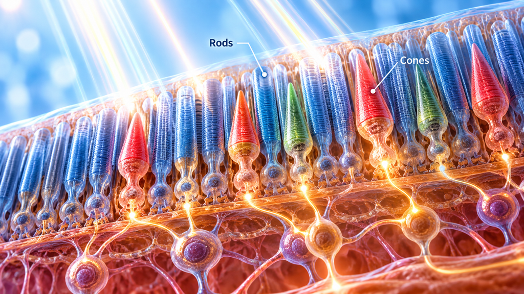
One of the most remarkable stages of vision occurs inside rods and cones.
When photons strike light-sensitive pigments within these cells, they trigger a series of biochemical reactions known as phototransduction.
In rod cells, the main visual pigment is called rhodopsin.
Light activates rhodopsin, initiating a molecular signaling cascade that changes the electrical state of the photoreceptor.
Interestingly, photoreceptors behave differently from many other sensory cells. They continuously release the neurotransmitter glutamate in darkness. When light activates them, they become more electrically negative — a process called hyperpolarization — and release less glutamate.
This change communicates information about incoming light to other retinal neurons.
How the retina processes visual signals
Photoreceptors communicate with bipolar cells, which pass information toward retinal ganglion cells.
Additional retinal neurons, including horizontal and amacrine cells, help modify these signals.
Together, these cellular networks contribute to the detection of contrast, brightness, and changes in the visual environment.
Ganglion cells then generate nerve impulses. Their long nerve fibers come together to form the optic nerve.
At this stage, light has been transformed into neural information that can travel toward the brain.
The Optic Nerve: Connecting Your Eyes to Your Brain
The optic nerve is a bundle of more than one million nerve fibers that carries visual information from the retina toward the brain. The journey does not end there.
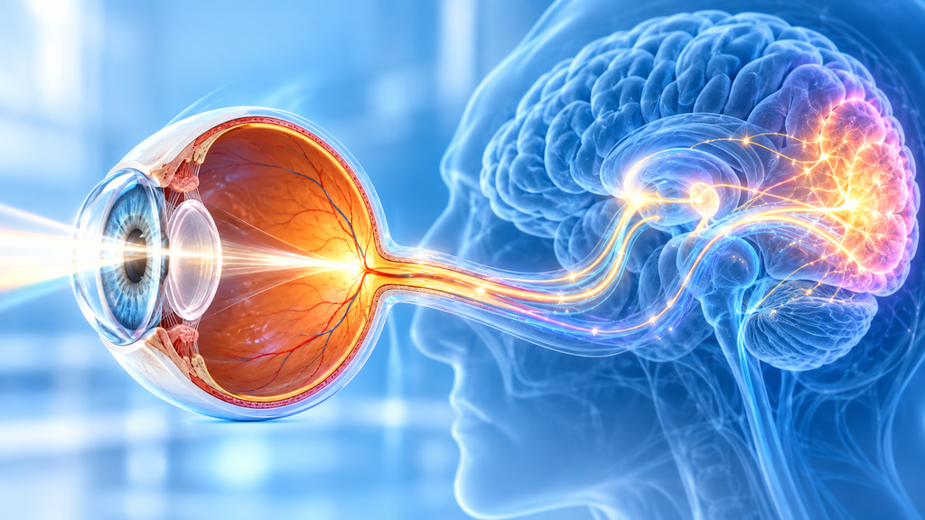
After the retina converts light into electrical signals, the optic nerve carries this information to the brain, where the visual cortex processes it into the images we perceive.
At a structure called the optic chiasm, some nerve fibers cross to the opposite side of the brain, while others remain on the same side.
This arrangement allows information from the right visual field of both eyes to be processed in the left hemisphere, and information from the left visual field to be processed in the right hemisphere.
Most visual signals then reach the lateral geniculate nucleus (LGN), a relay center within the thalamus.
From there, they travel through the optic radiations to the primary visual cortex in the occipital lobe, located at the back of the brain.
The visual cortex and its connected regions analyze information about shapes, edges, movement, color, and spatial relationships.
This coordinated processing ultimately contributes to the visual world we consciously experience.
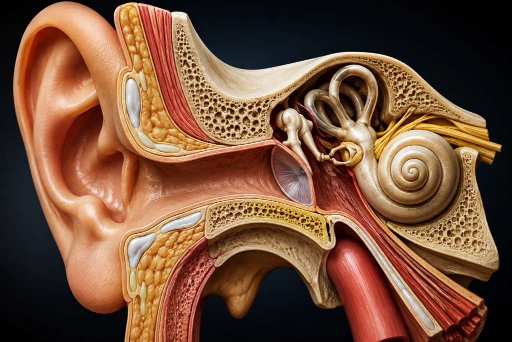
Similar sensory processing occurs in hearing, where sound waves are converted into electrical signals that the brain interprets as sound.
How Do We See Colors?
Your visual system continuously adjusts to changes in illumination.
When you enter a dark room after spending time outdoors, you may initially struggle to see. Gradually, your visual sensitivity increases as your pupils widen and your photoreceptors adapt.
Rods are particularly important during this process.
Conversely, moving from darkness into bright sunlight initially produces glare, followed by adaptation to the brighter environment.
These adjustments enable the visual system to operate across an enormous range of lighting conditions.
Why Can We See in Both Bright Light and Darkness?
Your visual system continuously adjusts to changes in illumination.
When you enter a dark room after spending time outdoors, you may initially struggle to see. Gradually, your visual sensitivity increases as your pupils widen and your photoreceptors adapt.
Rods are particularly important during this process.
Conversely, moving from darkness into bright sunlight initially produces glare, followed by adaptation to the brighter environment.
These adjustments enable the visual system to operate across an enormous range of lighting conditions.
How Do Both Eyes Create Depth Perception?
Because our eyes are positioned slightly apart, each receives a somewhat different view of the same object.
The brain compares these two perspectives through a process called stereopsis, helping us perceive three-dimensional depth.
This ability contributes to everyday activities such as catching a ball, pouring water into a glass, or judging distances while driving.
Depth perception also relies on other visual cues, including perspective, object size, motion, and shadows. Therefore, people can still perceive depth using only one eye, although some depth judgments become more difficult.
What Happens When the Visual System Stops Working Properly?
Clear vision depends on every stage of the visual pathway functioning correctly.
Different disorders can interfere with this process:
- Myopia (nearsightedness): Light focuses in front of the retina, making distant objects appear blurry.
- Hyperopia (farsightedness): Light would focus behind the retina when the eye is relaxed, often making nearby objects difficult to see clearly.
- Cataracts: Clouding of the lens causes blurry or hazy vision.
- Retinal disorders: Damage to photoreceptors or other retinal structures can interfere with the conversion and processing of light.
- Optic nerve damage: Disruption of the nerve fibers carrying visual information can produce permanent visual impairment.
Refractive errors can often be corrected with appropriate lenses, but conditions affecting the retina or optic nerve may require specialized treatment.
Frequently Asked Questions
Do we see with our eyes or our brain?
Both are essential. The eyes collect and process light, while the brain interprets the resulting neural signals to produce conscious visual perception.
Why do we have a blind spot?
The optic nerve leaves the retina at a region called the optic disc. This area contains no photoreceptors, creating a natural blind spot. Normally, your other eye and the brain’s processing help prevent you from noticing it.
Why is it difficult to see colors at night?
In dim light, vision increasingly relies on rods, which are highly light-sensitive but do not provide detailed color information. Cone-mediated color vision becomes less effective as illumination decreases.
Does the brain turn images upside down?
The cornea and lens produce an inverted optical image on the retina. However, the brain does not simply rotate a picture. It processes visual signals and constructs a stable perception of your surroundings.
Final Thoughts
Vision is much more than light entering the eyes. It is a sophisticated biological process involving precise optical focusing, specialized photoreceptors, complex retinal circuits, and advanced neural processing.
Every object you recognize and every color you perceive is the result of continuous communication between your eyes and your brain.
Understanding this process also explains why protecting your eyes, recognizing warning signs, and maintaining good eye health are essential for preserving vision. The process of how your eyes turn light into vision depends on precise cooperation between the cornea, lens, retina, optic nerve, and brain.
Sources
- How the Eyes Work | National Eye Institute — National Eye Institute.
- Phototransduction in Rods and Cones – Webvision – NCBI Bookshelf — NCBI Bookshelf, Webvision.
- Neuroanatomy, Visual Pathway – StatPearls – NCBI Bookshelf — NCBI Bookshelf.
- Types of Refractive Errors | National Eye Institute — National Eye Institute.
- Retinal Detachment | National Eye Institute — National Eye Institute.